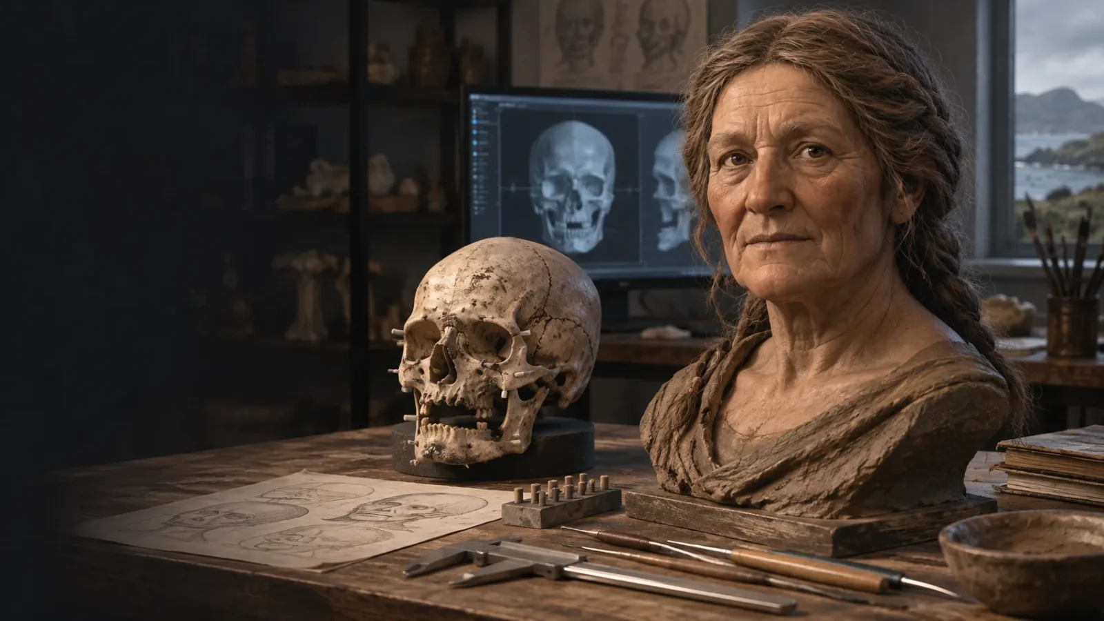
When a museum shows a reconstructed ancient face, the image can feel like a photograph from another time. Hilda’s face has that effect. She looks directly at us: an older woman with a lined brow, a bare mouth and an expression that seems to suggest a life behind it.
But the object behind the image is not a portrait. It is a skull, held by the University of Edinburgh and associated with a group of remains presented to the Edinburgh Phrenological Society in 1833. In 2019, forensic-art student Karen Fleming used a scan of the skull to make a three-dimensional wax reconstruction for a University of Dundee project.
The contrast tells us where the evidence ends. A skull can anchor a discussion of facial structure, while the finished approximation still depends on choices about soft tissue, hair, colour and expression.
Keep exploring
Continue with Curiosmos
Who was Hilda?
“Hilda” is a project nickname, not a name recovered from an Iron Age inscription. The skull was described in the nineteenth-century record as one of six “Druids of the Hebrides,” and the remains were associated with Stornoway on the Isle of Lewis. The University of Dundee’s account describes the woman as having been more than 60 years old when she died and says the skull was unable to be carbon-dated for the project.
The same account gives a broad date of about 55 BCE to 400 CE, based on information recorded in an 1833 journal. It is a reported range, not a new radiocarbon result. The surviving record stops short of a precise birth date, a personal name or a biography.
The word “druid” needs similar care. Dundee’s press release uses the term because of the historical label attached to the remains, while a skull alone offers no way to establish that its owner held a religious office. The reconstruction project gives us a face attached to a museum record and a set of historical claims—not a confirmed portrait of a known druid.
How is a face reconstructed from a skull?
Fleming’s reconstruction began with a three-dimensional scan. In the standard method described by forensic-art researchers, the skull is examined for features that affect facial proportions: the form of the jaw, the width of the orbits, the shape of the nasal opening, dental evidence, age-related changes and any asymmetry.
Markers representing average soft-tissue depths can then be placed at anatomical points. Muscles are built over the skull, followed by a layer representing skin and fat. The skull gives the practitioner a framework for the relationships between major features. Many surface details remain outside that record.
A review by Kathryn Lindsay, Frank Rühli and V. B. Deleon in The Anatomical Record explains why the process is best called facial approximation. Tissue-depth studies vary, measurements carry error, and the final stages—skin texture, hair, colour and some features of the eyes, nose and mouth—require judgement. Even a careful practitioner is working within a range of plausible faces.
What can the skull tell us?
The skull can constrain the broad architecture of a face. It can help estimate the proportions of the forehead, cheekbones, jaw and nose. Dental loss and changes to the bone can provide clues about age and health. Asymmetries in the skull may also be reflected in the reconstruction rather than smoothed away.
In Hilda’s case, Dundee described a toothless woman who had lost her teeth before death. That is a meaningful observation about her body. It can raise questions about diet, dental disease and the experience of ageing. From the dental evidence alone, we cannot reconstruct exactly what she ate, how she lost her teeth or what her daily life looked like.
The estimate that she lived beyond 60 is also easy to misread. An average life expectancy at birth is pulled down by infant and childhood deaths. That figure describes survival, not a rule that every adult was expected to follow. A long life may reflect health, social support or circumstances the surviving evidence cannot recover.
What does the reconstruction add?
The wax head turns measurements and a museum record into something most people can understand at a glance. That makes the method visible. Viewers can see that the face is built outward from bone, rather than painted onto an anonymous silhouette.
It also puts a person into a collection that was assembled through nineteenth-century ideas about race, character and the classification of human remains. The University of Edinburgh’s Anatomical Museum now describes a collection containing human skeletal remains alongside anthropology, phrenology, forensic and other material. That setting is part of Hilda’s modern history. The museum record is not neutral simply because the object is old.
The wax head performs two jobs at once. It can help explain forensic art, and it can prompt questions about how human remains were collected, named and displayed. Both uses remain valid even when the face is only a plausible likeness.
Which details are least certain?
The closer a detail is to the skin’s surface, the less securely the skull can determine it. Hair style, hair colour, eye colour, skin tone, scars and expression are not stored as a complete record in the bone. A reconstruction may choose a restrained appearance, but restraint is still a choice.
The same skull could produce noticeably different results if two artists made different decisions about tissue depth, eyelids, lips, hair or age-related soft tissue. That range is the point: the image is an interpretation built within anatomical limits.
For that reason, the image should be labelled a reconstruction or approximation. Calling it “what Hilda really looked like” turns a careful method into a promise the evidence cannot keep.
Can we know whether she was a druid?
The face alone offers no answer, and the skull is not a title or an inscription.
Druids are known through later Greek, Roman and Irish texts, and the term has often been applied too freely to Iron Age remains. A person buried in a particular place or with particular objects may support a discussion about ritual or status, but the label still needs archaeological context. The Hilda sources reviewed here establish a museum history and a reconstruction project; no direct inscription or burial record names her as a druid.
That does not make the story empty. It makes the strongest question more precise: how do researchers build a responsible image of a person when the surviving evidence is limited and the old labels are uncertain?
What the face adds to the record
Hilda’s reconstructed face visualises a set of constraints. The skull supports some proportions, the dental evidence records a history of tooth loss, and the museum record gives the object a documented modern trail. The finished face is one plausible result among several.
That boundary makes the story useful. A compelling image can invite us into the past, but the source record has to remain visible behind it. Hilda may have lived in the Iron Age, but the face looking back at us was made in a university studio in 2019.
For a related example of how archaeologists separate a striking object from the evidence around it, see Curiosmos’ guide to why archaeological context matters. The same discipline applies here: start with what the skull and records support, then mark the distance to everything that remains a possibility.
Sources and further reading
- University of Dundee, “Student reveals the face of Iron Age female druid”, 14 August 2019.
- University of Edinburgh, Anatomical Museum collection overview.
- K. E. Lindsay, F. J. Rühli and V. B. Deleon, “Revealing the Face of an Ancient Egyptian: Synthesis of Current and Traditional Approaches to Evidence-Based Facial Approximation”, The Anatomical Record, 2015.
- For comparison, see Curiosmos’ report on the Lady of Brassempouy and the limits of calling an ancient face a portrait.